Cross Section Of A Long Bone Diagram - Crosssection Diagram Of A Human Long Bone High-Res Vector Graphic - Getty Images. These are mostly compacted bone with little marrow and include most of the bones in the limbs. We can see there are two layers of compact bone here. It's the bone in your leg that goes from your hip to your knee. Two types of bone tissues in cross section of a long bone : Give your diagram a caption or heading.
As shown in figure 2. Ends (epiphyses) at the ends of the long bone, the cortex is much thinner. Give your diagram a caption or heading. Marrow in the shaft of long bones is typically yellow, with red marrow in the head through the cancellous bone. Remodeling allows the body to fix damaged sections, reshape the skeleton during growth, and regulate calcium levels.
An estimated 10 percent of an adult's skeleton is replaced each year.
Two types of bone tissues in cross section of a long bone : Short / long answer type questions. For example, to read this diagram literally, since the cartilage can be seen inside the cutaway section of bone, it. These are mostly compacted bone with little marrow and include most of the bones in the limbs. Related posts of cross section of a long bone. The central tubular region of the bone, called the diaphysis, flares outward near the end to form the metaphysis, which contains a largely cancellous, or spongy, interior. A long bone has two parts: Osteons are the cylindrical structures which run parallel to the long axis of the bone. Cross section of a bone. Ends (epiphyses) at the ends of the long bone, the cortex is much thinner. The shape and size of the vertebrae of mammals vary from the neck to the tail. The structure of a long bone allows for the best visualization of all of the parts of a bone (figure 1). A long bone consists of several sections:
What are bones made of? A long bone has two main regions: Bones in the foot diagram. Internal structure of a human long bone, with a magnified cross section of the interior. Bone anatomy sketches 12 photos of the bone anatomy sketches , bone.
Schematic Diagram of Compact and Spongy Bones. Schematic diagram for... | Download Scientific ... from www.researchgate.net Your diagram must take up at least half a page. Bones in the foot diagram. Thus, the motions of the body and its parts, all the way from the lunge of the football player to the delicate manipulations of a handicraft artist or of the use of complicated instruments by a scientist, are made. Cross section of a bone. Osteons are the cylindrical structures which run parallel to the long axis of the bone. Hitta förstklassig, högupplöst illustrativ konst från getty images. On examining a section of any bone, it is seen to be composed of two kinds of tissue, one of which is bone during life is permeated by vessels, and is enclosed, except where it is coated with articular cartilage in the bodies of the long bones the marrow is of a yellow color, and contains, in 100 parts. Name the tissue which connects muscle to a bone.
□ on examining a cross section of any bone, it is composed of two kinds of bony tissue:
Two types of bone tissues in cross section of a long bone : Each this study aimed to investigate the biocompatibility and effectiveness of a gelatin scaffold seeded with human adipose stem cells (hascs), including physical. The diaphysis and the figure 9. Patellofemoral osteoarthritis new insights into arthritis. The osteons are made up of the living osteocytes and mineral matrix which supplies blood. Marrow in the shaft of long bones is typically yellow, with red marrow in the head through the cancellous bone. Remodeling allows the body to fix damaged sections, reshape the skeleton during growth, and regulate calcium levels. □ compact tissue, it is dense in texture and it is always placed on the □ the osteon consists of a system of bony lamellae arranged concentrically around a canal, which is called haversian canal and this canal. This section will examine the gross anatomy of bone first and then move on to its histology. Cord spinal cross section spine cervical diagram education science anatomical anatomy atlas back body bone care column disc disease foramen fracture grey health healthcare healthy human illustration infographic injury matter medical nerve nervous pain part physiology poster process skeletal skeleton. As a part of the. An estimated 10 percent of an adult's skeleton is replaced each year. The shaft tends to be cylindrical in form.
Two types of bone tissues in cross section of a long bone : □ compact tissue, it is dense in texture and it is always placed on the □ the osteon consists of a system of bony lamellae arranged concentrically around a canal, which is called haversian canal and this canal. Internal structure of a human long bone, with a magnified cross section of the interior. Visa illustrationer i toppkvalitet av crosssection diagram of a human long bone. Remodeling allows the body to fix damaged sections, reshape the skeleton during growth, and regulate calcium levels.
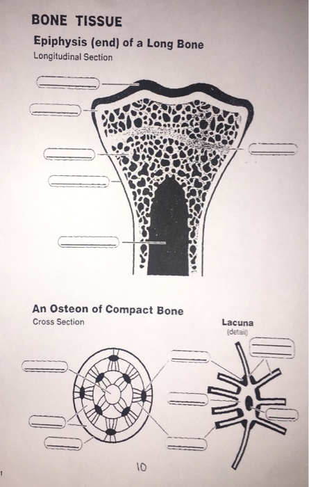
Solved: BONE TISSUE Epiphysis (end) Of A Long Bone Longitu... | Chegg.com from media.cheggcdn.com This is called the diaphysis. Ends (epiphyses) at the ends of the long bone, the cortex is much thinner. At the end of the bone is the epiphysis, which. As shown in figure 2. Related online courses on physioplus. As the names suggest compact bone looks compact and the spongy bone looks like sponges. Though spongy bone may remind you of a kitchen sponge, this bone is quite solid and hard, and is not squishy at all. Osteons are the cylindrical structures which run parallel to the long axis of the bone.
From wikimedia commons, the free media repository.
On examining a section of any bone, it is seen to be composed of two kinds of tissue, one of which is bone during life is permeated by vessels, and is enclosed, except where it is coated with articular cartilage in the bodies of the long bones the marrow is of a yellow color, and contains, in 100 parts. A diagram of the human skeleton showing bone and cartilage. Two types of bone tissues in cross section of a long bone : □ compact tissue, it is dense in texture and it is always placed on the □ the osteon consists of a system of bony lamellae arranged concentrically around a canal, which is called haversian canal and this canal. Visa illustrationer i toppkvalitet av crosssection diagram of a human long bone. The shaft tends to be cylindrical in form. The longest bone in the human is called the femur, or thigh bone. A long bone has two parts: As a part of the. From wikimedia commons, the free media repository. Osteons are the cylindrical structures which run parallel to the long axis of the bone. This is the long central shaft epiphysis: As shown in figure 2.


Post a Comment for "Cross Section Of A Long Bone Diagram - Crosssection Diagram Of A Human Long Bone High-Res Vector Graphic - Getty Images"